Figure 6.
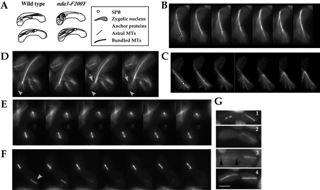
nda3-F200Y cells display persistent horsetail cortical contacts and show a high frequency of spindle failure in meiosis II. (A) Diagram of cortical contacts in horsetail movement. (B and C) Time-lapse single plane images of astral microtubules during horsetail movement in wild-type (B) and nda3-F200Y (AK101) (C) cells. An open circle in the first frame of each series indicates the position of the zygotic spindle pole body. (D) Detachment of an astral microtubule from the pole region while retaining cortical contacts was observed in both wild-type and nda3-F200Y cells. Arrowhead points to cortical contact; arrow indicates point of detachment. (E and F) Time-lapse images of asymmetric meiosis II spindle failure in nda3-F200Y cells. Arrows indicate unstable spindle of pair. (G) Early and late meiosis II phenotypes in nda3-F200Y cells showing 1) single failing bipolar spindle (left), 2) a single extended meiosis II spindle, 3) one robust and one weak (flanked by arrowheads) meiosis II spindle, and 4) normal meiosis II extended spindles. Bar for all images, 5 μm (shown in G).

