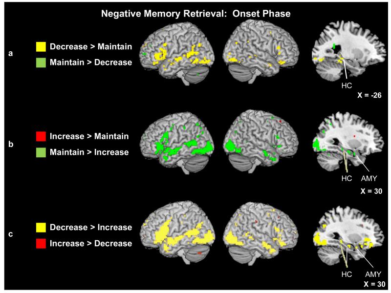
Figure 5.
(a) Neural activity for the decrease and maintain trials during negative AM onset. Saggital slice shows a region of left hippocampus (Tal: x = −26, y = −35, z = 0) that was more active during the decrease than maintain trials. (b) Neural activity for the increase and maintain trials during negative AM onset. Saggital slice shows a region of right amygdala (Tal: x = 30, y = −3, z = −17) as well as two regions of right hippocampus (Tal: x = 28, y = −14, z = −14; Tal: x = 30, y = −29, z = −7) that were more active during the maintain than increase trials. (c) Neural activity for the decrease and increase trials during negative AM onset. Saggital slice shows a region of right amygdala (Tal: x = 30, y = 1, z = −17) as well as two regions of right hippocampus (Tal: x = 26, y = −33, z = −8; Tal: x = 32, y = −16, z = −14) that were more active during the decrease than increase trials. Activity is significant at p < .001 and a 5-voxel threshold extent.

