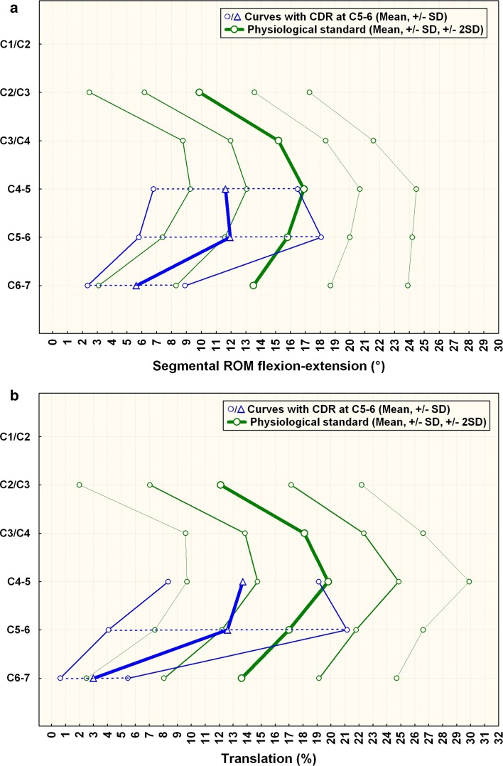
Fig. 4.
a Comparison of segmental ROM in patients with CDR at C5–6 compared to normals. C6–7 is the caudal-adjacent segment, C4–5 the supra-adjacent segment. The graph illustrates sagittal segmental rotation of C2–C7. The results of the CDR-group at the instrumented level, above and below are plotted against the physiological standard [10] ±1 SD (green smaller lines) and ±2 SD (green outer lines). b Comparison of segmental translation in patients with CDR at C5–6 (n = 12) compared to normals. C6–7 is the caudal adjacent segment, C4–5 the supra-adjacent segment. The graph illustrates sagittal segmental translation of C2–C7. The results of the CDR-group at the instrumented level, above and below are plotted against the physiological standard [10] ±1 SD (greensmaller lines) and ±2 SD (greenouter lines)

