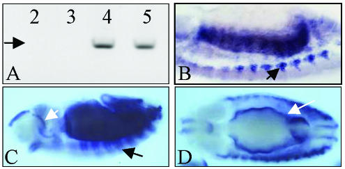
Figure 1.
wings-up A expression in the embryo. (A) RT-PCR of 2- to 5-h embryo extracts primed for TnI. Expression of a 1.2-kb cDNA (arrow) can be seen from stage 7 (4 h) onwards. Numbers indicate hours of development from 30 min. Egg-laying periods of the CS strain. (B) Lateral view of a 10-μm section of a stage 12 embryo revealing TnI transcripts in somatic muscle primordia (arrow) by in situ hybridization. (C) Lateral view of a whole mount stage 12 embryo showing TnI transcripts in segment arranged somatic developing muscles (black arrow), and in the foregut (white arrow). (D) Dorsal view of a whole mount stage 14 embryo showing TnI expression in developing visceral muscles (white arrow).

