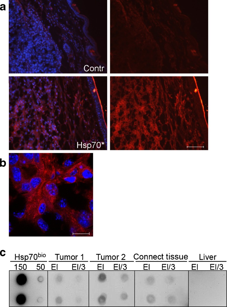
Fig. 1.
Migration of Hsp70 from gel into B16F10 tumor and subcutaneous tissue. a Morphological tracing of Hsp70 transport. Hsp70 was labeled with Alexa555 dye (Invitrogen, USA) according to instructions of the manufacturer and introduced in carbopol–glycerol–DMSO gel (700 μg of Hsp70 Alexa555 per milliliter of gel); 70 μl of gel was applied onto 7-day-old B16 tumor, and the sections of the tissue were prepared for the light microscopy. In the left part of Fig. 1, the pattern of staining of cell nuclei with DAPI is presented. Scale bar, 50 μm. b Microscopic analysis of Hsp70 transport into cancerous tissue. The sections were prepared as in A and studied with the aid of Leica TCS SP2 confocal microscope. Scale bar, 5 μm. c Biochemical proof of Hsp70 penetration inside B16 tumor. B16 tumor and adjacent tissue treated with gel-containing biotinylated Hsp70 were cut to obtain pieces with the approximate volume of 10–15 μl each. The proteins contained in each piece were extracted and subjected to the reaction with ATP–agarose gel; after washing, the proteins captured by the gel were applied on nitrocellulose, and spots were stained with neutravidin and biotinilated peroxidase solution. The dots are presented in pairs in the following order (from left to right): pure Hsp70 bio, 150 and 50 nanograms per dot; B16 tumor from animal no. 1, eluate 1:1(EL) and 1:3 (El/3); tumor from animal no. 2, eluate 1:1(EL) and 1:3(El/3); eluate obtained after processing of connective tissue adjacent to the tumor no. 2, eluate 1:1(EL) and 1:3(El/3); and eluate obtained after processing of liver of the animal no. 2, eluate 1:1(EL) and 1:3(El/3)

