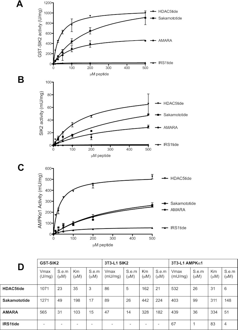
Figure 1. Identification of suitable peptide substrates for in vitro kinase activity measurements of SIK2.
(A) Purified wild-type or kinase-inactive (Lys49Met, not shown) GST–SIK2 (10 ng) expressed in HEK-293 cells was assayed in vitro using the following substrate concentrations: 0, 10, 25, 50, 100, 200 and 500 μM. (B and C) Endogenous SIK2 or AMPKα1 immunoprecipitated from 3T3-L1 adipocytes was used to evaluate the peptide substrates under the same conditions as for GST–SIK2. Results are means±S.D. for a triplicate assay. (D) Non-linear regression using a Michaelis–Menten model was used for determining Vmax and Km values using GraphPad Prism 5.

