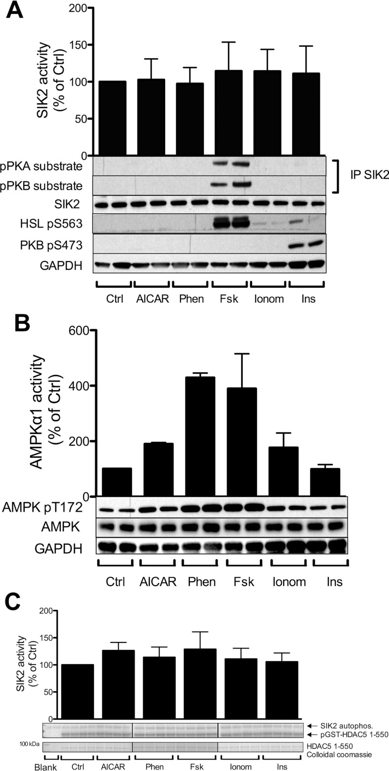
Figure 2. Phosphorylation and activity of SIK2 in 3T3-L1 adipocytes treated with various cellular stimuli, including cAMP inducers.
Fully differentiated 3T3-L1 adipocytes were treated with one of the following agents: AICAR (2 mM, 1 h), phenformin (1 mM, 1 h; Phen), forskolin (50 μM, 15 min; Fsk), ionomycin (1 μM, 5 min; Ionom) or insulin (100 nM, 10 min; Ins). (A) Lysates were analysed with regard to immunoprecipitated endogenous SIK2 activity in vitro towards the peptide substrate HDAC5tide and phosphorylation by PKA and PKB employing consensus motif (pPKA and pPKB substrate) antibodies. PKB phosphoSer473 and HSL phosphoSer563 were used as controls. Activity data presented are means±S.D. from four to six individual experiments. Blots shown are representative of 5–14 experiments. (B) Lysates were analysed with regard to immunoprecipitated endogenous AMPKα1 activity in vitro towards the peptide substrate AMARA, and phosphorylation of the T-loop using anti-phosphoThr172 antibodies. (C) Lysates were analysed with regard to immunoprecipitated SIK2 activity in vitro towards HDAC5 (1–550) purified protein by autoradiography as described in the Experimental section. Activity data are presented as means±S.D. from three individual experiments. Ctrl, control.

