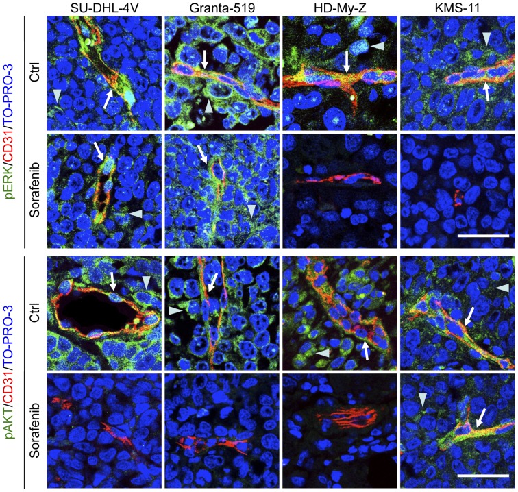
Figure 7. Sorafenib-induced inhibition of Akt and ERK phosphorylation in tumor and endothelial cells.
SU-DHL-4V, Granta-519, HD-MyZ and KMS-11 tumor nodules growing subcutaneously in mice treated with sorafenib (90 mg/kg) or control vehicle (DMSO) for 5 days were excised 3 hours after the last treatment, fixed in formalin and embedded in paraffin. Tumor sections were double-stained with CD31 (red) and phospho-ERK 1/2 (green) or phospho-Akt (green) followed by the appropriate AlexaFluor 568- or 488-conjugated secondary antibody for indirect immunofluorescent detection of the corresponding antigen. Nuclei were detected with TO-PRO-3 nuclear dye (blue). Arrows indicate phospho-ERK 1/2 or pospho-Akt expression by endothelial cells; arrowheads indicate phospho-ERK 1/2 or pospho-Akt expression by tumor cells. Representative images are shown. Objective lens, original magnification: 1.0 NA oil objective, 40×. Scale bar: 50 µm.

