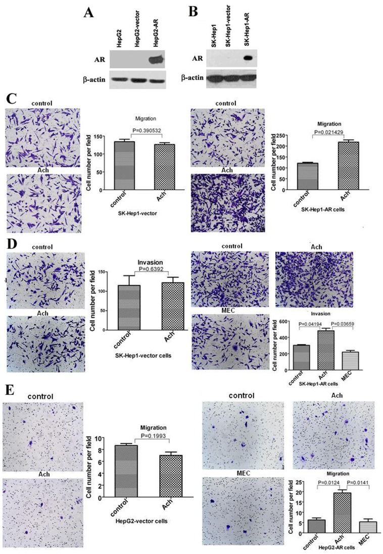
Figure 3. The effects of Ach on migration and invasion of AR negative SK-Hep1 cells and HepG2 cells, SK-Hep1-AR cells, and HepG2-AR cells.
(A, B) Western blots revealed expression of AR in HepG2-AR and SK-Hep1-AR cells. HepG2 and SK-Hep1 cells were infected by the lentivirus vector as a control or the lentivirus expressing AR. β-actin served as a loading control of total proteins. (C–D) AR negative SK-Hep1 cells infected with the lentiviral-AR (SK-Hep1-AR) were analyzed for migration (C) and invasion (D) by transwell cell migration and invasion assays after treatment with Ach for 24 h and 48 h respectively. SK-Hep1 cells infected with the lentiviral-empty-vector (SK-Hep1-V) served as a control. (E) AR negative HepG2 cells infected with the lentiviral-AR (HepG2-AR) were analyzed for migration by after treatment with Ach for 24 h. HepG2 cells infected with the lentiviral-empty-vector (HepG2-V) served as a control. “*” indicated statistically significant differences (p<0.05).

