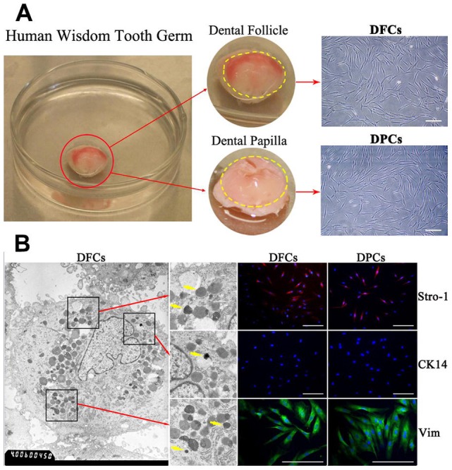
Figure 1. Culture and identification of DFCs and DPCs.

(A) Primary DFCs and DPCs from human wisdom tooth germ. The dental follicle on the crown surface has visible blood supply. The dental papilla located inside the crown is transparent and jelly-like. Both DFCs and DPCs show typical fibroblast-like spindle morphology under a light microscope. (B) Homogeneous electron-dense granules in DFCs indicated by yellow arrows are featured for DFCs. Both cells were positive for STRO-1 and for Vimentin, but negative for epithelial marker CK-14. Scale bar = 100 µm in (A) and (B).
