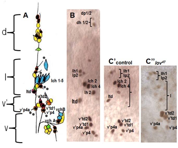
Figure 6. Lov expression in the embryonic abdominal PNS.
A - a cartoon of the four clusters (dorsal (d), lateral (l), ventral’ (v′) and ventral (v)) of sensory neurons within each hemisegment of the abdominal segments (redrawn from [2]). Neurons are color coded according to class as follows: orange - external sense organ, pink - tracheal dendrite, blue - chordotonal, yellow - dendritic arborization, green - bipolar dendritic. Key neurons discussed in the text are labeled. Neuronal nuclei expressing Lov (shown by black dots or ovals) were identified in embryos co-stained with anti-Lov and 22C10. Grey dots = putative non-neuronal support cell nuclei that express low levels of Lov. Nomenclature for all neurons as in [24] except that the neurons of the lateral chordotonal organ are labeled lch1-5. B - Actual example of wild type Lov-staining nuclei at stage 15 in clusters d, l, and v′ of a single abdominal hemisegment. C - anti-Lov (brown) and 22C10 (blue) costaining of the lateral chordotonal organ in a control embryo to demonstrate Lov limitation to lch2 and lch4. D - 22C10 staining of a lov47 homozygous embryo demonstrating the presence of all five chordotonal neurons in the lateral chordotonal organ. E′, E′′ - loss of Lov staining in chordotonal organ neurons lch2 and 4 of a lov47 mutant embryo. E′ shows control staining in clusters l and v′. E′′ shows staining in equivalent regions of a lov47 embryo. Loss of staining in td neuron ltd is also seen in this lov47 embryo.

