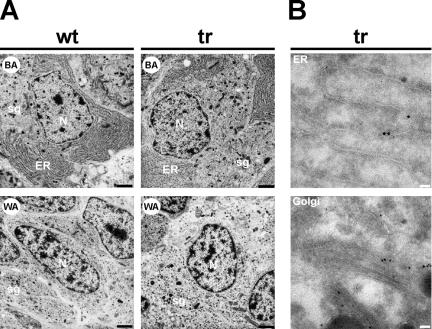
Figure 3.
Electron microscopy on transgenic Xenopus intermediate pituitary cells. (A) Electron micrographs of melanotrope cells of wild-type (wt) and #124 transgenic (tr) Xenopus adapted to a black (BA) or white (WA) background. N, nucleus; ER, endoplasmic reticulum; sg, secretory/storage granule. Bar, 2 μm. (B) Pituitary glands from transgenic (tr) frogs (F1 #224, expressing high levels of Xp24δ2-GFP) were subjected to immunoelectron microscopical analysis. For immunodetection, the anti-GFP antibody was used in combination with protein-A-gold to visualize the Xp24δ2-GFP fusion protein. Immunoreactivity was found in structures that resemble the ER and the Golgi. Bars, 0.1 μm.

