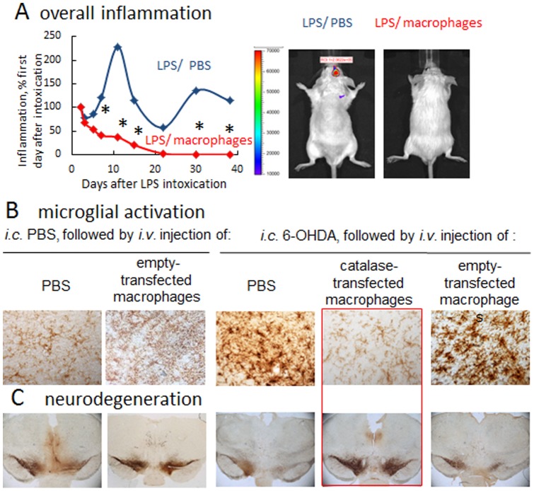
Figure 3. Anti-inflammatory and neuroprotective effects of catalase-transfected macrophages in PD murine models.
A: LPS-induced encephalitis in BALB/C mice were injected i.v. with catalase-transfected macrophages (red curve), or PBS (blue curve). IVIS images over 40 days were taken ten minutes after intraperitoneal (i.p.) injection of a XenoLight RediJect probe for inflammation. The chemiluminescent signal was quantified and presented as radiance ratios of treated animal after 24 hours after LPS injection and at various times thereafter. Genetically-modified macrophages caused prolonged decreases of neuroinflammation in LPS-intoxicated mice. IVIS representative images at day 30 are shown. (B) and (C): BALB/c mice were i.c. injected with 6-OHDA. Forty eight hours later animals were i.v. injected with catalase-transfected macrophages, and 21 days later they were sacrificed, and mid-brain slides were stained for expression of B: CD11b, a marker for activated microglia or C: TH, a marker for dopaminergic neurons. Whereas 6-OHDA treatment caused significant microglia activation and neuronal loss, administration of catalase-transfected macrophages dramatically decreased oxidative stress, and increased neuronal survival. Administration of empty-vector transfected macrophages did not affect microglia activation, or number of dopaminergic neurons in in mice with brain inflammation. Statistical significance (shown by asterisk: p<0.05) was assessed by a standard t-test compared to mice with i.c. LPS injections followed by i.v. PBS injections (healthy controls). Values are means ± SEM (N = 4).

