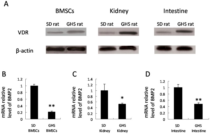
Figure 1. VDR levels and BMP2 expressions in GHS and SD rats.
Nuclear proteins were isolated from BMSCs and tissues of SD and GHS rats and were subjected to Western blot using anti VDR. The representative data are shown in (A). Expressions of BMP2 in BMSCs, kidney and intestine tissues from GHS rats decrease relative to SD rats (B, C, D). (*, P<0.05; **, P<0.01).

