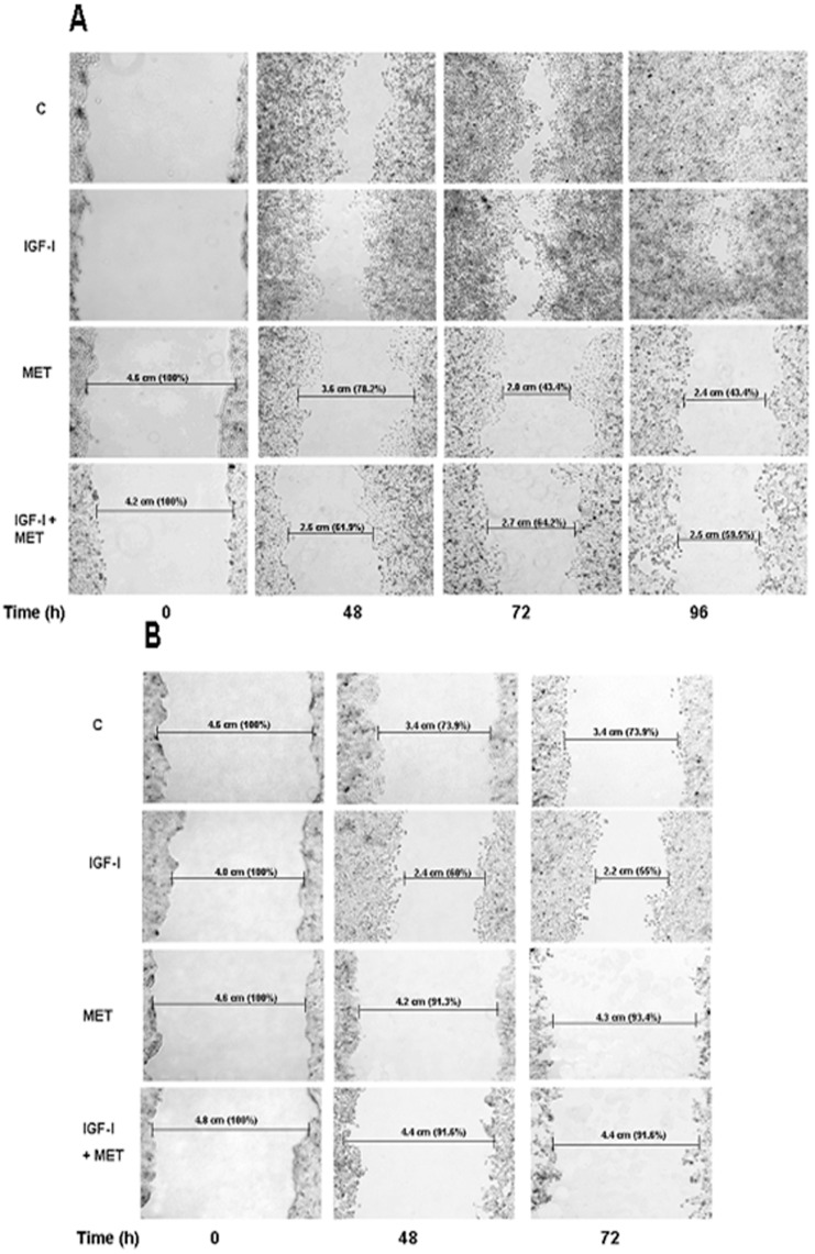
Figure 5. Effect of metformin on cell migration.
Wounds were made on monolayers of USPC-2 (A) and USPC-1 (B) cells grown to 100% confluence. Cells were then incubated in serum-free media containing IGF-I (50 ng/ml), metformin (10 mM), or both, for 48, 72 and 96 h (USPC-2) and for 48 and 72 h (USPC-1). Treated or untreated (control) cells were photographed just after scratch (time 0), and after 48, 72 and 96 h. Results presented here are representative of triplicate independent samples of each cell line. The rate of migration was measured by quantifying the total distance that the cells (as indicated by rulers) moved from the edge of the scratch toward the centre of the scratch. A value of 100% was given to the wound area at time 0. The migration of IGF-I and/or metformin treated samples was compared to wound area at time 0.

