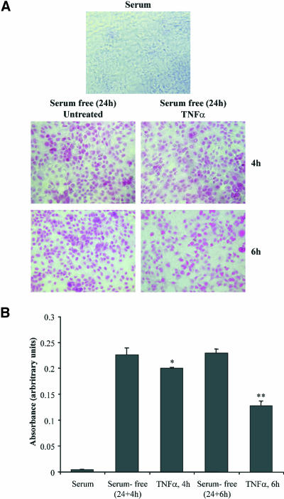
Figure 1.
Antiapoptotic effects of TNF-α in OK cells. Cells in complete medium or cells that had been exposed to serum-free medium for 24 h and then incubated with TNF-α for the indicated times were assessed for apoptosis using the colorimetric APOPercentage apoptosis assay. Apoptotic cells were photographed using an inverted microscope (A) or quantified by measuring the absorbance by using a microplate colorimeter (B). Mean + SEM from two separate experiments performed in triplicate (significance level *p < 0.05, **p < 0.01).

