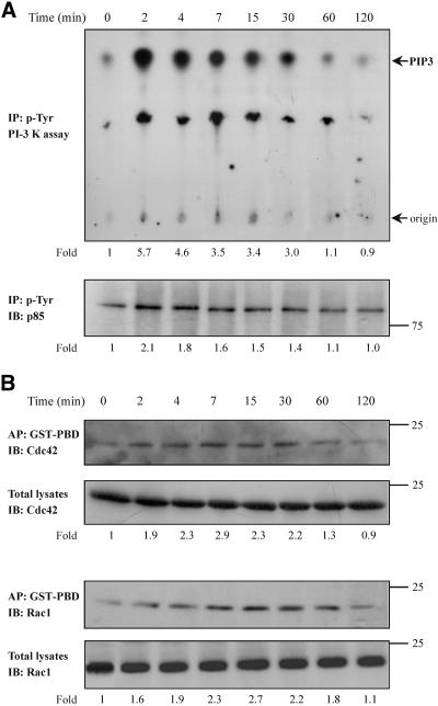
Figure 4.
Effect of TNF-α on the PI-3 kinase and Cdc42/Rac1 activity. Cells were incubated for the indicated times with TNF-α (10 ng/ml). Equal amount of proteins of cell lysates were immunoprecipitated with an anti-phosphotyrosine antibody and subjected to an in vitro PI-3 kinase assay, as described in MATERIALS AND METHODS, by using PIP2 as substrate. The number below each lane indicates the fold amount of phosphatidylinositol-3,4,5-trisphosphate (PIP3) product, with that of untreated cells taken as 1 (top). The phosphorylation of the p85 regulatory subunit of PI-3 kinase that was immunoprecipitated in the kinase assay was assessed by immunoprecipitation (IP) with an anti-phosphotyrosine antibody and immunoblotting (IB) with anti-PI-3 kinase (p85) antibody. The number below each lane indicates the fold phosphorylation of p85, with that of untreated cells taken as 1 (bottom). (B) Equal volume of cell lysates from untreated or TNF-α-treated cells were affinity precipitated (AP) with GTP-PBD bound to glutathione-agarose beads. Precipitated GTP-Cdc42 or GTP-Rac1 was detected by IB with anti-Cdc42 or anti-Rac1 antibody, respectively. Equal volume of total lysates from untreated and TNF-α-treated cells were subjected to SDS-PAGE, transferred to nitrocellulose membrane, and IB with monoclonal anti-Cdc42 or anti-Rac1 antibody, respectively. The number below each lane indicates the normalized fold activation of Cdc42 or Rac1, with that of untreated cells taken as 1. Results shown are representative of three independent experiments with similar results.

