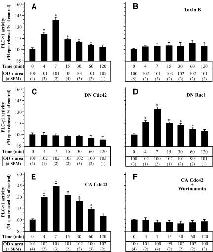
Figure 7.
PLC-γ1 activity depends on Cdc42 activation in TNF-α-treated cells. PLC-γ1 was immunoprecipitated from equal amount of proteins from TNF-α-treated cells (A) or cells that had been preincubated with toxin B (B) or had been transfected with Cdc42(T17N) (C), Rac1(T17N) (D), Cdc42V12 (E), or with Cdc42V12 followed by exposure to wortmannin (100 nM, 30 min) (F) and then incubated with TNF-α for the indicated times. PLC-γ1 was analyzed for hydrolytic activity toward [3H]phosphatidylinositol 4,5-bisphosphate as described in MATERIALS AND METHODS. PLC-γ1 activity is presented as percentage of control cells' activity by measuring the cpm [3H]inositol 1,4,5-triphosphate released (mean + SEM from three separate experiments performed in duplicate, *p < 0.05). To confirm that equal amounts of PLC-γ1 protein were assayed under each condition separate immunoblot experiments were performed. The “OD × area” (as percentage of control) (±SEM) corresponding to bands of PLC-γ1 loaded was measured by PC-based image analysis and presented in table below each panel.

