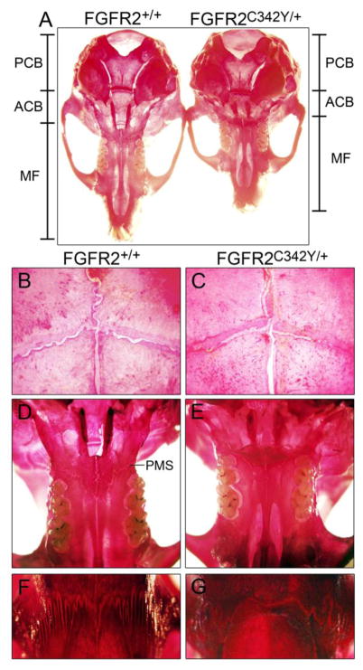
Fig. 3. Skeletal stains of BALB/c FGFR2C342Y/+ and FGFR2+/+ calvaria.
(A) Stained whole calvaria from four week old FGFR2C342Y/+ mice and wild type littermates demonstrates that the anterior cranial base (ACB) and midface (MF) appear shorter in FGFR2C342Y/+ mice, while the posterior cranial base (PCB) appears relatively unaffected. (B,C) Visualization of the coronal sutures at 32x magnification shows increased frontal to parietal bone overlap with potential partial fusions in FGFR2C342Y/+ mice (b) compared to wild type mice (c). (D,E) Visualization of the inferior aspect of the midface at 6.3x magnification shows apparent fusion of the palatomaxillary suture (PMS) in FGFR2C342Y/+ mice (d) but not in FGFR2+/+ mice (e). (F,G) Visualization of the nasofrontal suture at 32x magnification shows a lack of interdigitation with points of apparent fusion between the frontal and nasal bones in FGFR2C342Y/+ mice (f) but not in FGFR2+/+ mice (g).

