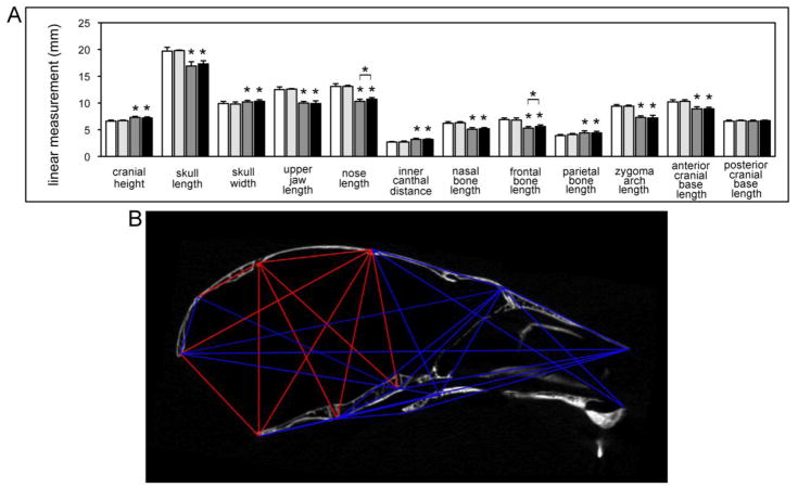
Fig. 6. Linear and morphometric analysis of craniofacial forms demonstrate consistent and craniofacial bone specific skeletal abnormalities in BALB/c FGFR2C342Y/+ mice.
(A) Three-dimensional coordinate data generated from landmarks placed micro CT scans of FGFR2C342Y/+ and FGFR2+/+ mice were used to generate linear measurements (Fig. 1). Measurements of craniofacial bones and cranial vault dimensions demonstrate significantly diminished skull length, upper jaw length, nasal bone length, frontal bone length and zygomatic arch length with significantly increased cranial height, cranial width, inner canthal distance and parietal bone length in FGFR2C342Y/+ as compared to wild type mice, regardless of gender. Nose length and frontal bone length were significantly larger in male FGFR2C342Y/+ as compared to female FGFR2C342Y/+ mice but no other gender differences were found. Data is presented as means +/− standard deviations. Statistical significance was established by the student’s t test. *p < .001 vs. wild type or between indicated groups. White = FGFR2+/+ female, light grey = FGFR2+/+ male, dark grey = FGFR2C342Y/+ female, black = FGFR2C342Y/+ male. (B) A representative subset of landmark distance EDMA mean ratios is shown on a sagittal micro CT section of an FGFR2C342Y/+ mouse. Blue lines indicate those distances that are significantly smaller in FGFR2C342Y/+ as compared to FGFR2+/+ mice. Red lines indicate those distances that are significantly larger in FGFR2C342Y/+ as compared to FGFR2+/+ mice. All distances are for midline landmarks other than those for landmarks 8,9 and 10,11. Distance ratios for bilateral landmarks are represented as a single line and were significant for both right and left sides. Note that larger distances in the FGFR2C342Y/+ mouse are primarily restricted to the parietal bones, interparietal bones and posterior cranial base dimensions.

