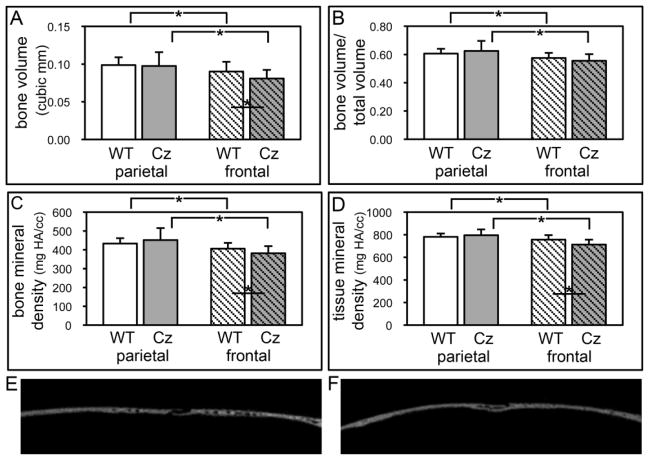
Fig. 7. Diminished bone volume and density in frontal bones of BALB/c FGFR2C342Y/+ mice.
(A,B,C,D) Micro CT analyses demonstrate significantly diminished bone volume, bone mineral density and tissue mineral density in frontal bones but not parietal bones of FGFR2C342Y/+ (Cz) mice as compared to wild type (WT) mice. Bone volume, bone volume/total volume, bone mineral density and tissue mineral density are also significantly diminished in frontal compared to parietal bones of both FGFR2C342Y/+ and wild type mice. *p<.05. (E,F) Wild type (E) and FGFR2C342Y/+ (F) sagittal sections of parietal (left) and frontal (right) bones surrounding the coronal suture.

