Figure 4.
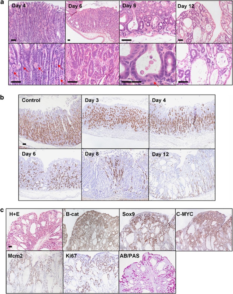
Activation of WNT signalling in the corpus of the stomach leads to FGPs with progressive parietal cell loss. (a) H+E stain of corpus region from AhCreER+ GSK3 alphafl/fl betafl/fl (GSK3) mice at days 4, 6 and 12 post-induction (top, low magnification; bottom, high magnification). Day 4—areas of the corpus show a reduction in the number of parietal cells (red arrows). Day 6—both FGPs and regions of adenomatous change are present in the corpus (red arrow marks parietal cells lining a fundic cyst). Day 12—widespread fundic gland polyposis and adenomatous change in the corpus. Scale bar, 50 μm. (b) IHC detecting H+–K+–ATPase expression marking parietal cells in AhCreER+ GSK3 alphafl/fl betafl/fl versus AhCreER+ (Control) mice at days 4, 6, 8 and 12 post-induction. Scale bar, 50 μm. (c) FGPs and adenomatous change in AhCreER+ GSK3 alphafl/fl betafl/fl mice at day 12 post-induction. IHC staining for β-catenin, c-MYC, Sox9, Mcm2, ki67 and Alcian Blue Periodic Acid Schiff (AB/PAS). Scale bar, 50 μm.

