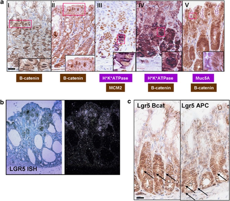
Figure 5.
Recombination occurs within multiple cell types in the corpus. (a) IHC detecting β-catenin showing nuclear localisation in parietal cells (I) and foveolar cells (II) from AhCreER+ APCfl/fl mice at day 3 post-induction. Double IHC staining detecting Mcm2 (brown) and H+K+ATPase (violet) in parietal cells (III). β-Catenin (brown) and H+K+ATPase (violet) in parietal cells (IV). β-Catenin (brown) and Muc5A (violet) in foveolar cells (V). Scale bar, 10 μm. (b) ISH for Lgr5 in corpus tissue from AhCreER+ GSK3 alphafl/fl betafl/fl mice at day 12 post-induction. Bright-field (left) and dark-field microscopy (right) images. (c) IHC detecting nuclear β-catenin in Lgr5creERT2+ Catnbexon3fl/exon3fl (Lgr5Bcat) and Lgr5creERT2+ APCfl/fl (Lgr5 APC) mice at day 28 and 50 post-induction, respectively. Arrows indicate cells with nuclear β-catenin that have not progressed. Scale bar, 20 μm.

