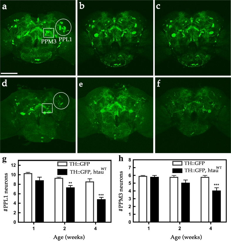
Fig. 1.
Expression of htauWT induces age-dependent DA neuron demise. Representative confocal images show mCD8-GFP-marked DA neurons in age-matched control fly brains (a–c TH::mCD8-GFP) and htauWT-expressing brains (d–f TH::htauWT, mCD8-GFP) of one- (a, d), two- (b, e), and four- (c, f) week-old individuals. Two clusters of DA neurons, PPL1 (protocerebral posterior lateral 1, circles) and PPM3 (protocerebral posterior medial 3, squares), are indicated. Scale bar 100 μm. Cell counts of PPL1 (g) and PPM3 (h) DA neurons from age-matched control and htauWT brains. Values shown represent Mean ± SEM (unpaired t test, **P < 0.001 at week 2 for g; ***P < 0.0001 at week 4 for g and h; n = 12)

