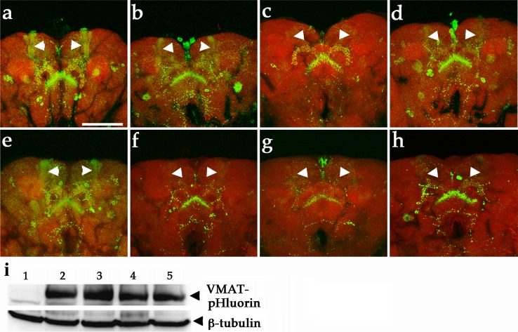
Fig. 6.
DA neurons expressing htauWT cause early impairment of vesicular dopamine release to nerve terminals as visualized by VMAT-pHluorin. a–f Confocal images of DA neurons from brains of 1-day-old (a–d) and 3-week-old (e–h) flies show VMAT-pHluorin reporter (green). a DA neurons expressing VMAT-pHluorin (TH::VMAT-pHluorin) reveal localization of VMAT-pHluorin to presynaptic terminals in brain regions, and b age-matched DA neurons co-expressed VMAT-pHluorin and htauWT (TH::VMAT-pHluorin, UAS-htauWT) show decreased GFP signals in mushroom bodies (arrowheads). e A representative confocal image shows the VMAT-pHluorin signals to mushroom bodies and other brain structure remains prominent (green) in 3-week-old control brain, while the GFP signals localized to mushroom bodies is diminished in DA neurons expressing htauWT (f, arrowheads). c, g Expression of htauAP evokes severe loss of pHluorin signaling compared to age-matched tauWT (b, f) and TauE14 (d, h). Rhodamine-phalloidin (red) marks the brain structure. Scale bar 100 μm. i Representative western blot shows the expression levels of VMAT-pHlurion of 1-week-old fly brains in control and experiment groups. 1 TH-GAL4, 2 TH::VMAT-pHluorin, 3 TH::VMAT-pHluorin, htauWT, 4 TH::VMAT-pHluorin, htauAP, 5 TH::VMAT-pHluorin, htauE14. Anti-β-tubulin served as a loading control

