Figure 4.
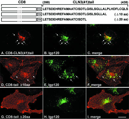
CD8-CLN3tail chimeras in NRK cells. CD8-CLN3(41)tail chimeras were expressed in stably transfected NRK cells and localized confocal microscopy after fixation and permeabilization with methanol and double labeling with antibodies to CD8α (A, D, and G) and lgp120 (B, E, and H). The CD8-CLN3(41)tail chimera (A) colocalized significantly (see arrows and merged images in C) with lpg120 in lysosomes (B). Removal of 10 amino acids from the C-terminus of the chimera (D) resulted in partial colocalization with lgp120 (E), but a significant fraction was seen at the cell surface. Lysosomal targeting of the chimera was completely lost by deletion of 20 amino acids (G) resulting in most of the protein being on the cell surface and essentially no overlap with lgp120 (H). Merged images of D/E and G/H are shown in F and I, respectively. Bar, 10 μm.

