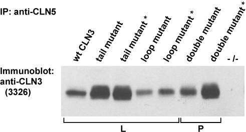
Figure 6.
Physical interaction between CLN5 and CLN3 in COS-1 cells. COS-1 cells were transiently transfected with either the full-length or C-terminally truncated CLN5 carrying SWE mutation (asterisk) and wt CLN3 or CLN3 carrying mutations either in the loop [LI(253-4)AA] or tail (M409A+G419A) targeting motifs or both (double mutant). Cell lysates were immunoprecipitated with CLN5 specific antibody, followed by immunoblotting with CLN3 specific antiserum (3326). -/- lane corresponds to nontransfected control cells. The observed differences in the strength of signal were directly proportional to expression levels of the different CLN3 constructs (our unpublished data). L and P indicate primary localization of CLN3 proteins in lysosomes and at the plasma membrane, respectively, in transfected HeLa cells (see Figures 1 and 5).

