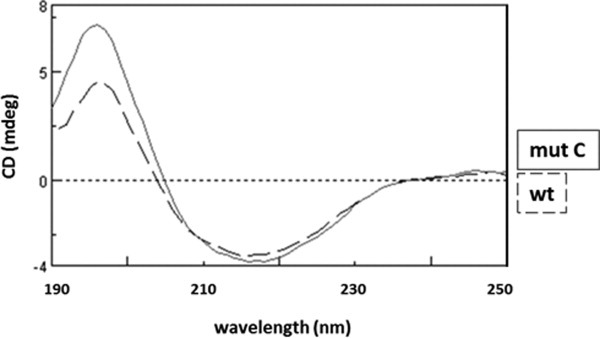
Figure 4.

CD spectra of refolded wild-type Txnip and Txnip mutant C. The wt protein and mutant C (mut C) were refolded, extensively dialyzed against a buffer containing 20 mM Tris–sulfuric acid and 0.5 M sodium fluoride (pH 7.5), and then concentrated to 0.5–1 mg/ml using an Amicon ultra-4 concentrator with a 10,000 molecular weight cut-off. Spectra were recorded between 190 and 260 nm with a 1 nm pitch at a 50 nm/min scanning speed using a JASCO J-815 CD spectrometer. The discontinuous line is the spectra for wt Txnip and the solid line is the spectra for mut C.
