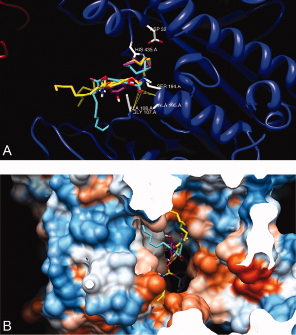
Figure 12.

Superimpositions of structures of tridentate 1 (yellow), bidentate 5 (turquoise), and monodentate 9 (mangenta) that have been automatically docked into the X-ray crystal of CEase 1AQL6 by AutoDock program.41, 44–46 View from the active site (A) and from the entrance (mouth) (B) of the enzyme. [Color figure can be viewed in the online issue, which is available at wileyonlinelibrary.com.]
