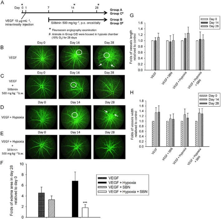
Figure 6.
Silibinin prevents VEGF- and VEGF plus hypoxia-induced retinal oedema and neovascularization in a rat AMD model. (A) Schematic of the VEGF-induced and VEGF plus hypoxia-induced retinal angiogenesis models in the BN rat. Fluorescein angiography was performed on day 0, 14 and 28. (B) In the group receiving intravitreal VEGF, the treated eye revealed diffusion of fluorescein from the vessel, representing increased permeability of retinal tissues (indicated by circle) and retinal oedema (indicated by arrow). (C) Silibinin treatment (500 mg kg−1 on days 7–28) decreased the VEGF-induced oedema, retinal permeability, and microvascular angiogenesis. (D) Animals injected with VEGF and exposed to hypoxia (10% O2) for 28 days show synergistic effects on retinal oedema (indicated by circle and arrow). The diameter and length of the retinal vessels were obviously increased. (E) Silibinin treatment of animals injected with VEGF and exposed to hypoxia (10% O2) for 28 days exhibit delayed and diminished AMD-related microvascular angiogenesis and oedema. The quantification of the day0, day14 and day 28 vessel length, vessel width and the oedema area were shown in Figure 6F, G and H. Data are expressed as mean ± SE from 5 independent experiments. ***P < 0.001 compared with the day 0 as control groups.

