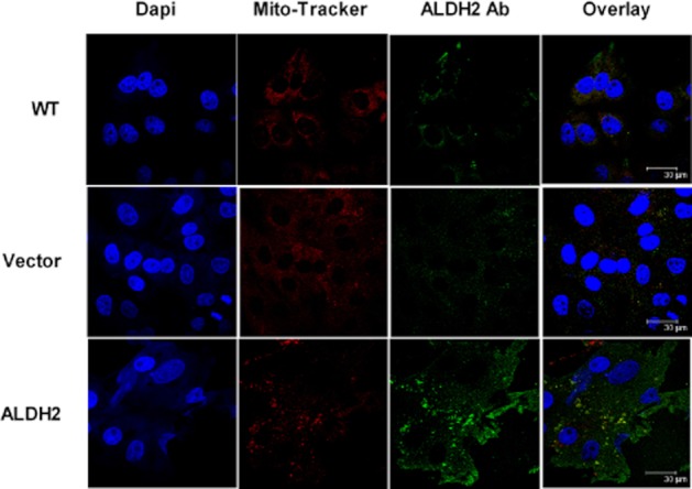
Figure 2.

Subcellular location of ALDH2 in PK1 cells. Cells grown on coverslips were fixed with 4% formalin. Nuclei were visualized by staining with DAPI, mitochondria by staining with MitoTracker and ALDH2 by labelling with goat anti-rabbit antibody conjugated with Alexa Fluor 546, after incubation with an antibody directed against ALDH2. Images were obtained by confocal microscopy. Note the increased expression of ALDH2 in PK1ALDH2 cells, both in the cytosol, and co-localized with MitoTracker.
