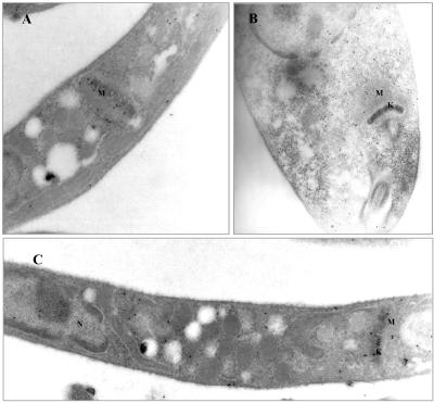
Figure 6.
Immunoelectron microscopy localization of HMGR in L. major promastigotes overexpressing HMGR1 and HMGR2. Cells were fixed in 4% paraformaldehyde and 0.1% glutaraldehyde and embedded in LRWhite resin. Thin sections were immunolabeled with anti-LmHMGR (dilution 1:70) polyclonal antibodies followed by goat anti-rabbit immunoglobulin conjugated to gold (10-nm probe). Magnification is 25,000× in A and C and 31,500× in B. (A) Section of L. major promastigotes transfected with the expression vector pSP72hmgr1 expressing a form of HMGR that lacks the first 14 amino acids. (B and C) Longitudinal sections of L. major promastigote cells transfected with the expression vector pSP72hmgr2 expressing a form of HMGR that lacks the first 19 amino acids. Increased gold labeling of the cytoplasm is evidenced. G, glycosome; K, kinetoplast; M, mitochondrion.

