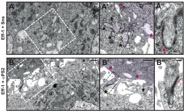
Fig. 4. Invasive finger-like membrane protrusions promote fusogenic protein engagement.

(A-A″) Electron micrographs of Sns-Eff-1-expressing cells. Boxed area in (A) enlarged in (A′), and that in (A′) enlarged in (A″). (A) A low magnification view of two adherent cells. (A′) Cell on the right (pseudo-colored purple) extended a group of finger-like protrusions (black arrows) to invade the cell on the left. Segments of electron-dense materials were present on the membranes along the protrusive fingers, but absent elsewhere on the cell membrane (magenta arrows). (A″) At a higher magnification, the electron-dense materials appeared like ladders between the two apposing cell membranes (red arrowheads). (B-B″) Electron micrographs of αPS2-Eff-1-expressing cells. Boxed area in (B) enlarged in (B′), and that in (B′) enlarged in (B″). (B) A low magnification view of the two adherent cells. (B′) Cell on the top (pseudo-colored purple) extended individual finger-like protrusions to invade the cell at the bottom. The invasive fingers (black arrows) were scattered along the broad cell-cell contact zone, and were associated with segments of electron-dense materials, which were absent elsewhere on the cell membranes (magenta arrows). (B″) At a higher magnification, the electron-dense materials also showed a ladder-like appearance (red arrowheads) between the two apposing membranes, as in Sns-Eff-1-expressing cells (A″). Scale bar: 500 nm (A, A′, B, and B′); 100 nm (A″ and B″).
