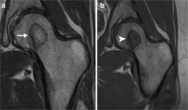
Fig. 8.
Femoral lesion in patient with breast cancer, suboptimally evaluated on proton density images. a Coronal proton density MR image of the left hip demonstrates a low-signal circle in the left femoral head, surrounding a region that has signal similar to that of surrounding marrow (arrow). The findings could be consistent with avascular necrosis. b Corresponding coronal T- weighted MR image clearly shows that the lesion has signal intensity similar to that of muscle (arrowhead), indicating that it represents a marrow-replacing lesion. Biopsy results confirmed metastatic adenocarcinoma of the breast

