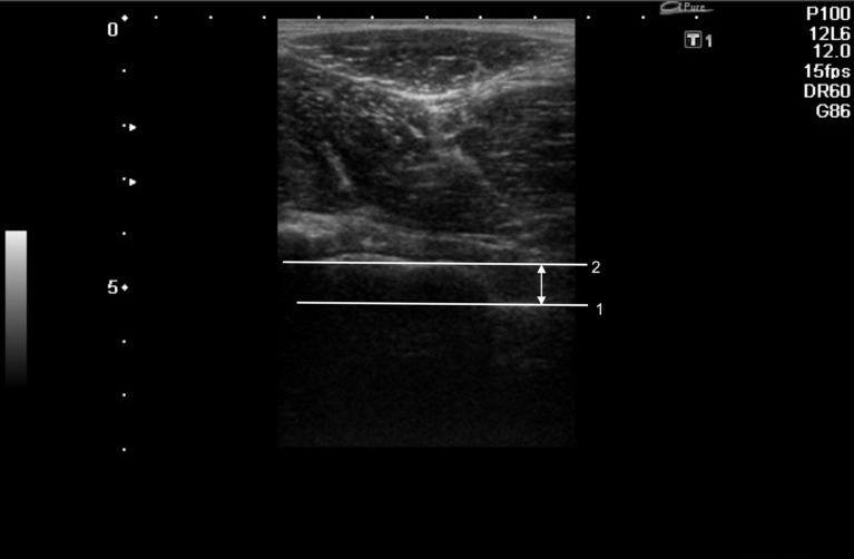
Fig. 3.
Ultrasound of the hip joint in 20° internal rotation. The anterior femoral distance is measured as the distance between the two lines. The first line (1) is placed as a tangent to the visible surface of the femoral neck; the second line (2) is drawn parallel to the first along the greatest perpendicular depth of epiphyseal overgrowth at the anterior femoral head-neck junction

