
Figure 1.
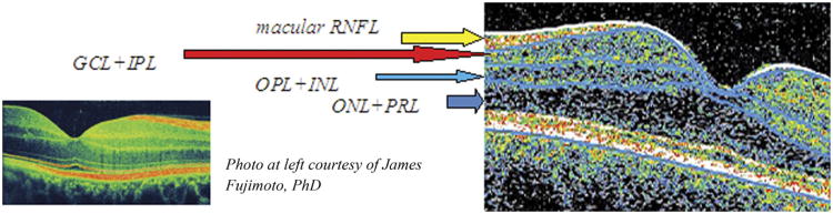
Vertical spectral-domain optical coherence tomography (OCT) section of right eye macula illustrating retinal segmentation techniques. The software algorithm delineates 4 macular layers: (1) Macular retinal nerve fiber layer (RNFL); (2) ganglion cell layer and inner plexiform layer (GCL+IPL); (3) outer plexiform layer and inner nuclear layer (OPL+INL); and (4) outer nuclear layer and photoreceptor layer (ONL+PRL). The software also calculates segmental thickness in 3 concentric circles with diameters of 1, 3, and 6 mm divided into equal quadrants in a diagonal orientation, similar to the pattern provided in commercial OCT.