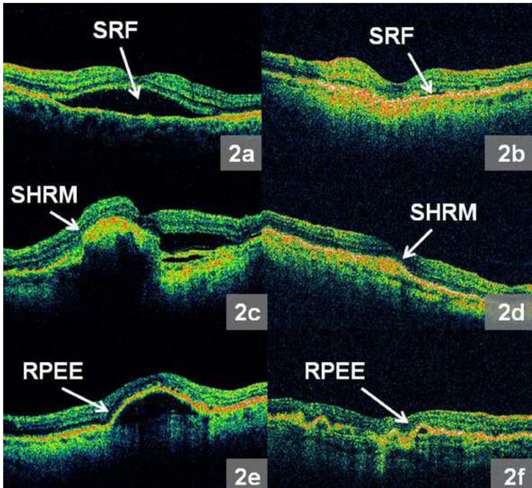
Figure 2.
a – f: Representative morphologic features from Optical Coherence Tomography (OCT) images produced by the macular thickness map (MTM) protocol: 2a. Obvious subretinal fluid (SRF), 2b. Subtle SRF, 2c. Obvious subretinal hyperreflective material (SHRM), 2d. Subtle SHRM, 2e. Obvious retinal pigment epithelium elevation (RPEE), 2f. Subtle RPEE.

