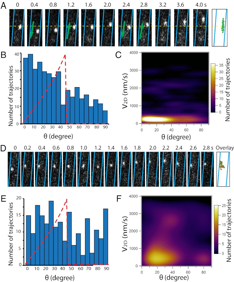
Fig. 7.
Helical motility of AglR is disrupted in the ΔagmU and ΔaglZ backgrounds. (A) AglR moves in linear trajectories in the ΔagmU strain, with frequent pauses and reversals. (B) Angle distribution of AglR motility deviates significantly from that of WT (dotted lines) at θ < 30°. (C) Projected V2D of motile molecules is slow and shows no correlation with θ. (D) AglR molecules move actively along irregular trajectories in the ΔaglZ strain. (E) Angle distribution of AglR motility (n = 203) deviates significantly from that of WT (dotted lines) in the ΔaglZ strain. (F) V2D-max of AglR in the ΔaglZ strain is higher than in the WT. AglR slowed down at random positions, evidenced by the irrelevancy between V2D and θ. (Scale bars, 1 µm.) Only sections of cells are shown in this figure. The images of cells with at least one cell pole in sight are shown in Movies S14 and S15.

