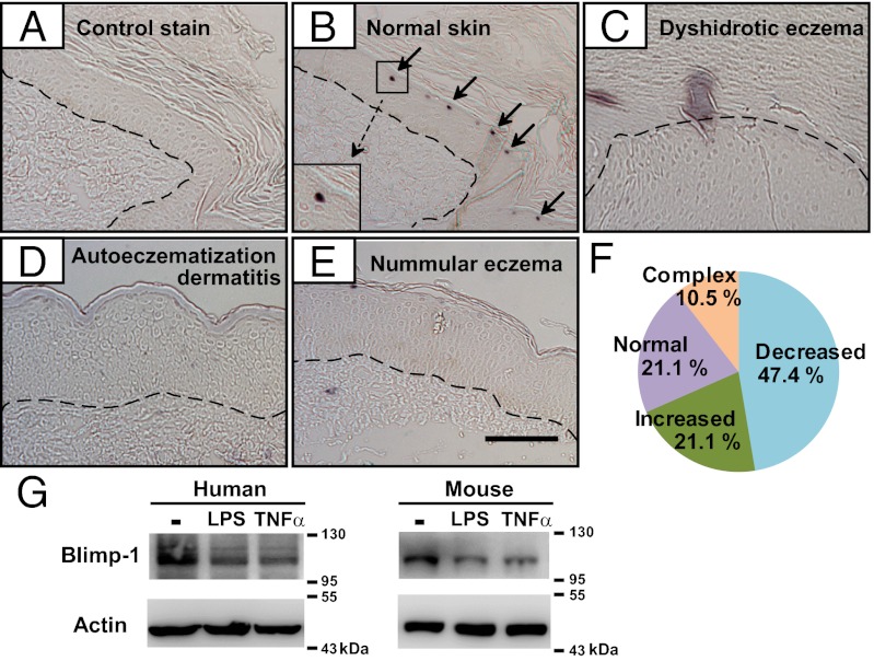
Fig. 1.
Expression of Blimp-1 in human epidermis and stimulated keratinocytes. (A–E) Immunohistochemical staining of human skin sections with isotype control antibody, mouse IgG (A) or with anti–Blimp-1 (B–E) or from individuals with normal skin (A and B), dyshidrotic eczema (C), autoeczematization dermatitis (D), or nummular eczema (E). Each dashed line denotes the epidermis–dermis boundary. Arrows indicate Blimp-1+ cells. (Scale bar: 100 μm.) (F) Relative expression of Blimp-1 from unclassified eczema skin samples (n = 19) compared with normal skin. Eczema skin sections that contained areas of both increased and decreased Blimp-1 were categorized as complex. (G) Immunoblots showing reduced Blimp-1 protein in human (Left) and mouse (Right) primary keratinocytes treated with LPS (5 μg/mL) or TNF-α (50 ng/mL), or untreated (−), for 24 h; results are representative of three experiments.

