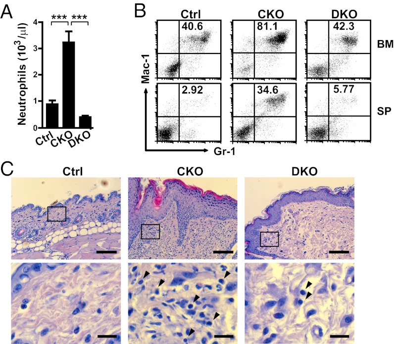
Fig. 5.
G-CSF contributes to inflammatory diseases in CKO mice. (A) The number of neutrophils in peripheral blood of the indicated mice at 5–6 mo after 4OHT injection was measured with an automated hematology analyzer. Data are mean ± SEM (n = 4). ***P < 0.005. (B) Populations of Gr-1+Mac-1+ neutrophils in BM and splenocytes (SP) of the indicated mice shown in a representative dot plot from two independent experiments. (C) H&E staining of neck skin from the indicated mice at 6 mo after 4OHT injection. The enlarged images in the boxed areas of the upper panels are shown in the lower panels. Arrowheads indicate lymphocytes. (Scale bars: 100 μm in upper panels; 15 μm in lower panels.)

