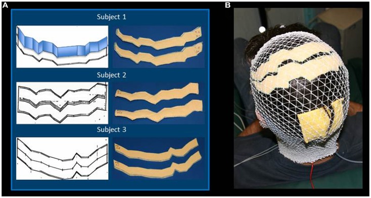
Figure 1.
Regional Personalized Electrode shaping. The sponge electrodes are shaped, for each subject, in two steps: first, drawing the left and right central sulci on a piece of paper using SoftTaxic software from a volumetric MRI; second, fitting the central sulcus by 2 cm-width parallelograms (an example is reported for Subject 1). For each S1 and M1 electrode, the shape is than drawn on and cut out of two sponge sheets; the two sheets are sewed together to allow the insertion of conductive material (A). The stimulating electrodes are positioned by proper neuronavigation procedure (reference landmark is visible frontally), while the reference electrode is positioned according to the alignment used by Feurra et al. (2011a). Electrodes are secured through an elastic cotton cap (B).

