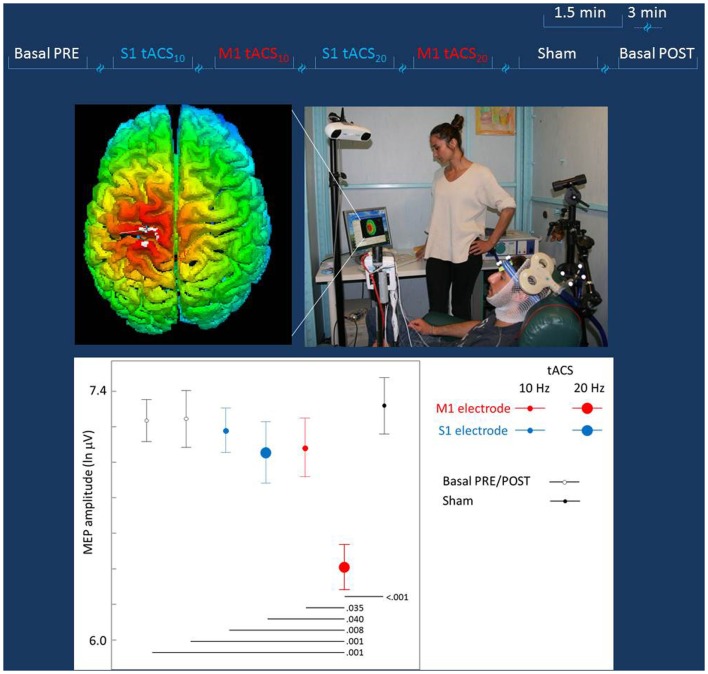
Figure 2.
Experiment to probe differential effects of stimulation target. Top lines: representation of the stimulation blocks, lasting 1.5 min each and intermingled by more than 3 min. The blocks (Sham, S1, or M1 tACS at 10 or 20 Hz) were randomly delivered across subjects. Middle Left: real time scalp projection of the TMS coil position onto the 3D-rendered cortical surface overlying the tCS electrodes to probe online the different effects on the cortical excitability as induced by the stimulation of the two cortical targets (the cross indicated the center of the coil over the central sulcus). Middle Right: experimental setup of the TMS session. The TMS focal coil overlies the tCS personalized M1 electrode; the TMS coil position is stabilized by a mechanical arm that is digitized once the OP hot-spot is identified and monitored throughout the experiment duration. Bottom: the MEP amplitude in the different tCS conditions. Post hoc comparisons are reported for significant differences.

