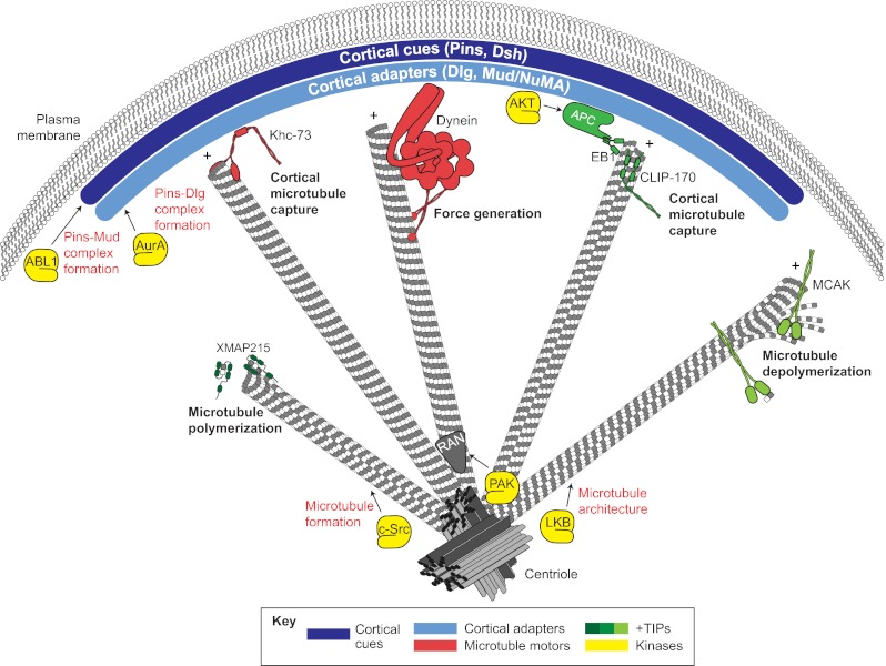
Fig. 4.
A summary of the mechanisms that contribute to spindle orientation. Cortical spindle orientation cues, such as Pins and Dsh (dark blue), polarize to specific regions of the cell cortex. Cortical adapters, such as Dlg and Mud/NuMA (light blue), are then recruited by direct interactions with cortical cues. Polarized cortical cues and adapters interface with spindle microtubules through interactions with various plus-end localized microtubule-binding proteins, including motor proteins (e.g. Khc-73 and Dynein; red) and additional regulatory complexes (e.g. EB1-APC; green). These elements provide microtubule capture and forge generation effects that coordinate spindle positioning. Additional plus-end binding proteins aid in microtubule dynamics, such as polymerases (e.g. XMAP215, dark green) and depolymerases (e.g. MCAK, light green), which are necessary for proper spindle assembly and function. An assortment of protein kinases (yellow) have been shown to regulate spindle orientation through various mechanisms, including regulation of microtubule architecture (e.g. LKB and c-Src), regulation of cortical cue-adapter complexes (e.g. ABL1 and AurA) and localization of microtubule regulators such as Ran (gray) (e.g. PAK).

