
Fig. 6.
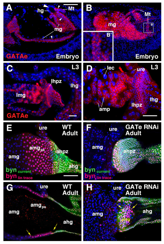
Role of GATAe in the formation of the posterior terminal midgut. (A-D) Confocal sections of gut of late embryo (A,B) and third instar larva (C,D), showing expression of GATAe mRNA (red). Embryonic expression is found in the midgut (mg; small arrowheads) and Malpighian tubules (Mt) (B′), but not in the hindgut (hg; big arrowhead). In the larva, GATAe is expressed strongly in adult midgut progenitors (amp) and the hindgut proliferation zone (hpz); faint expression is seen in the Malpighian tubules and ureter (ure), and differentiated midgut enterocytes (lec). (E-H) Confocal z-projection images of the adult hindgut-midgut boundary of wild-type (E,G) and GATAe knock-down animals (F,H). Surface views (E,F) and sagittal views (G,H) are shown. Cells of the hindgut proliferation zone are lineage traced from late third instar using byn-Gal4 driver. Current expression of byn is shown in green, the lineage is shown in red. In wild type, HPZ-derived cells come to lie within the posterior terminal midgut (amgps; arrows in G). Upon GATAe knock-down, HPZ-derived cells do not epithelialize and form a solid cluster of cells (plug; arrows) in the lumen of the hindgut (F,H). ahg, adult hindgut; ahpz, adult hindgut proliferation zone; amg, adult midgut; amgps, posterior terminal midgut; amp, adult midgut progenitor; hg, hindgut; lec, larval enterocyte; lhg, larval hindgut; lhpz, larval hindgut proliferation zone; lmg, larval midgut; mg, midgut; Mt, Malpighian tubule; ure, ureter. Scale bars: 50 μm.