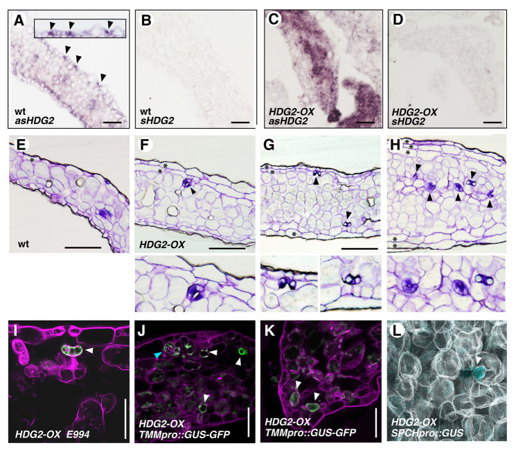
Fig. 2.
Ectopic overexpression of HDG2 confers ectopic stomata within mesophyll tissues and formation of multiple epidermal layers. (A-D) In situ hybridization analysis of HDG2 expression in 10-day-old wild-type (A,B) and HDG2-OX (C,D) seedlings treated with HDG2 antisense (A,C) or sense (B,D) probes. In wild type, endogenous HDG2 shows stomatal-lineage expression (A, arrowheads). Inset shows an enlarged image. Scale bars: 40 μm. (E-H) Histological cross-sections of plastic-embedded 10-day-old cotyledons from wild-type (E) and transgenic plants overexpressing HDG2 (HDG2-OX), which are CaMV35S::HDG2 (F-H). In HDG2-OX, ectopic differentiation of stomata in internal tissues (arrowheads) is evident. Bottom insets indicate enlarged images. Multiple epidermal layers (asterisks) occasionally formed. Images are taken under the same magnification. Scale bars: 100 μm. (I-K) Live sections of HDG2-OX cotyledons expressing mature guard cell marker E994 (I) and stomatal-lineage marker TMMpro::GUS-GFP (J,K). GFP is detected in ectopic internal stomata (I, arrowhead) and internal stomatal-lineage cells (J,K, arrowheads). A mature ectopic internal stoma is also evident in J (cyan arrowhead). Scale bars: 20 μm. (L) Optical section of mPS-PI stained mesophyll layer from HDG2-OX cotyledon expressing SPCHpro::GUS (arrowhead).

