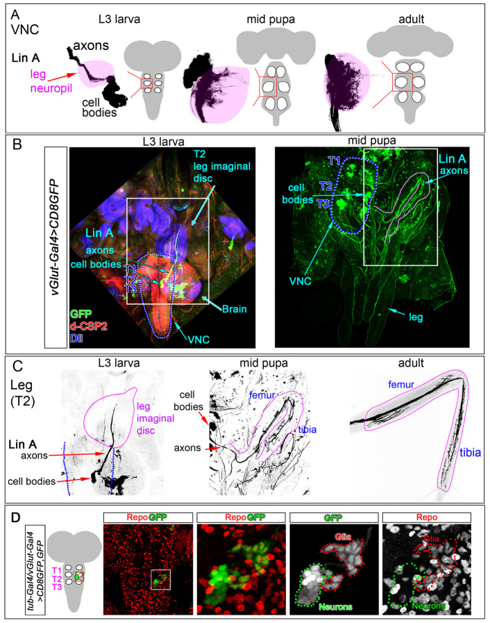
Fig. 1.
LinA MNs during development. (A-C) LinA MARCM clones in T2, labeled with vGlut-Gal4 >CD8GFP. (A) Cell bodies and dendrites of LinA MNs at late L3, mid-pupa and adult stages. Leg neuropil region is shaded pink. (B) LinA MARCM clones stained for d-CSP2 (red, labels the neuropil) and Dll (blue, labels the leg imaginal discs). VNCs are outlined with blue dotted lines. Regions shown in C are marked with white boxes. The pupal stage leg is outlined in pink. (C) LinA MN axons in the periphery. Shown is the GFP channel from the regions boxed in B. Late L3 leg imaginal disc, pupa stage leg and adult leg are outlined in pink. The VNC at the L3 stage is outlined with blue dotted lines. (D) LinA MARCM clones stained with anti-Repo (red) to mark glia. The left-most image shows a low-magnification view of the VNC (as depicted in the schematic); the region boxed is shown at higher magnification in the images to the right. About 22 LinA-derived glia are visualized; these surround the neuropil in the adult (data not shown).

