Fig. 1.

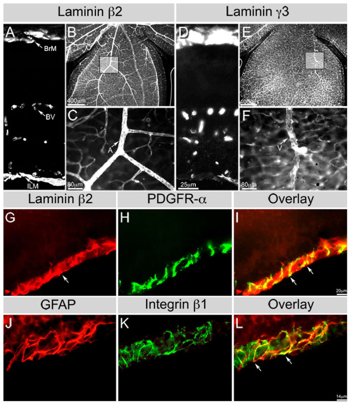
Astrocytes migrate within β2-containing laminin-rich niche. (A-C) Laminin β2 distribution in the wild-type P15 retina (A, section; B,C, wholemount). Boxed area in B is shown in C. β2 chain is deposited in all retinal basement membranes (BMs). (D-F) Laminin γ3 distribution in the wild-type P15 retina (D, section; E,F, wholemount). Boxed area in E is shown in F. The γ3 chain is deposited most prominently in microvessel and venous BMs; some deposition is seen in the inner limiting membrane (ILM). Scale bar in D applies to A,D. BrM, Bruch’s membrane; BV, blood vessels. (G,H) Wild-type P0 retinal section reacted with antibodies against laminin β2 chain (G) and PDGFRα (H). (I) Merge of G and H. Arrow in G indicates laminin β2 chain at the ILM. Arrows in I indicate PDGFRα-labeled astrocytes interacting with β2 chain-containing laminins at the ILM. (J,K) Wild-type P0 retinal section reacted with antibodies against GFAP (J) and β1 integrin (K) antibodies. (L) Merge of J and K. Arrows in L indicate β1 integrin in GFAP+ astrocytes.
