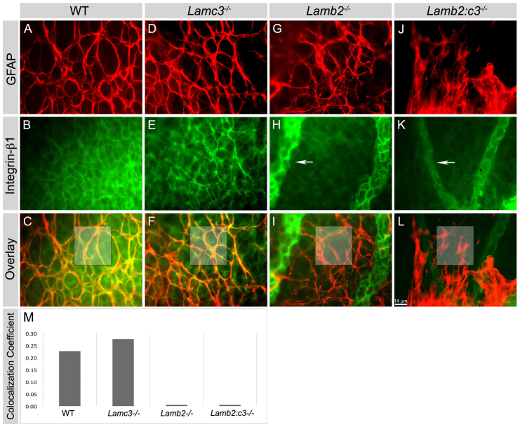
Fig. 4.
Deletion of laminins affects expression of β1 integrin in astrocytes in vivo. (A-L) The expression of integrin β1 was studied in P5 retinal wholemounts. GFAP+ astrocytes (red) express integrin β1 (green) in both wild-type and Lamc3-/- (yellow, C and F) retina but not in the Lamb2 mutants (I and L). Integrin β1 expression in endothelial cells (GFAP- cells) in all genotypes, particularly in the persistent hyaloid vessels (arrows, H and K). (M) Pearson’s colocalization coefficient was calculated for the boxed areas in images C-L. Scale bar: 14 μm.

