Fig. 5.
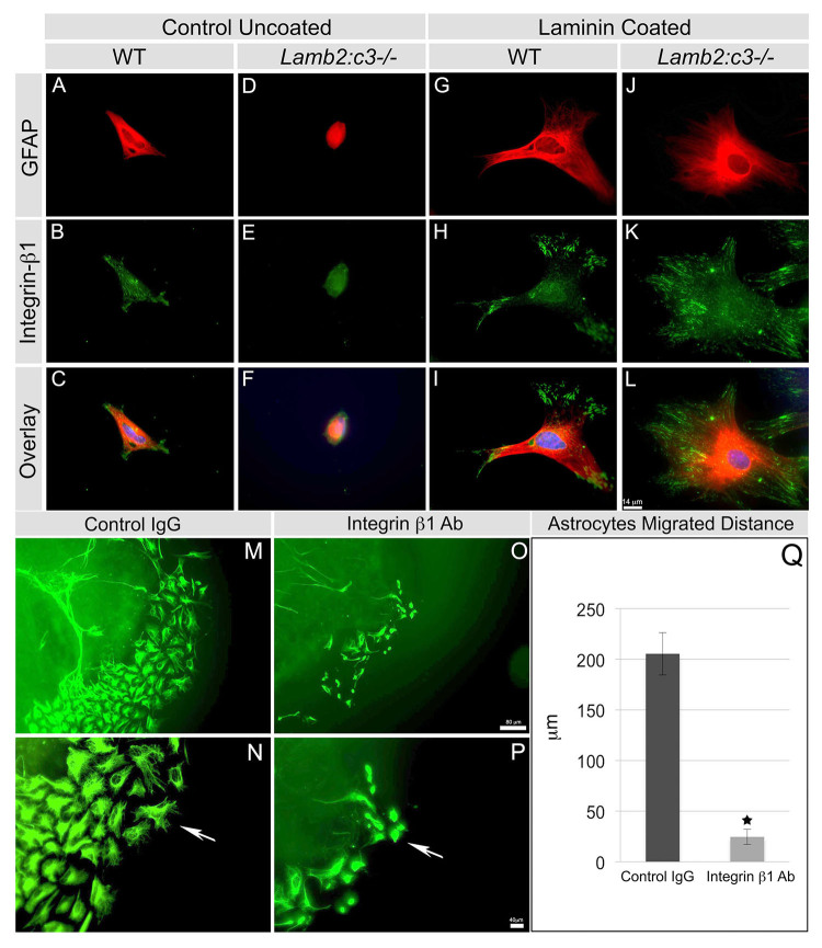
Exogenous laminin restores focal localization of β1 integrin in laminin-null astrocytes. (A-F) The astrocyte structure from P2 wild-type (A-C) and mutant (D-F) retinas were analyzed after 24 hours of culture with the indicated markers. DAPI (blue) labels the nucleus in the overlays. (G-L) EHS-laminin-coated substrates enhanced the spread of P2 wild-type (G-I) and mutant (J-L) astrocytes. (M-P) Function-blocking antibodies against integrin β1 prevent astrocyte migration from ONH explants. Explants were grown on EHS-laminin substrates in the presence of control IgG (M,N) or integrin β1 antibody (O,P) for 48 hours. EHS-laminin induced robust astrocyte (GFAP+) migration in the control treatment (M) and migrating cells showed elongated filopodia (arrow, N). Treatment with integrin β1 antibody reduced astrocyte migration (O) and filopodial elongation (P). Scale bars: 14 μm in A-L; 80 μm in M,O; 40 μm in N,P. (Q) Quantification of distance that astrocytes migrated away from the explants. Error bars represent s.e.m. *P<0.01.

