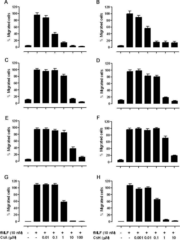
Figure 3. Effects of cyclosporins on fMLF-stimulated chemotaxis in FPR1 haplotype-expressing CHO-Gα16 cells.
(A and B) CH1, (C and D) CH2, (E and F) CH3 and (G and H) CH5. Cells were pre-incubated with different concentrations of cyclosporins for 15 min before loading into the upper wells of a 48-well chemotaxis chamber with 10 nM fMLF placed in the lower wells. The chemotaxis assay was conducted at 37°C for 4 h, and the numbers of migrated cells were determined by analysis with the Image-Pro Plus software, i.e. counting the integrated absorbance of cells after staining with 0.01% Crystal Violet in 20% methanol. Three fields were photographed for each well. The results presented are means±S.E.M. from three independent experiments, each in triplicate, showing the percentage of maximal chemotaxis induced with fMLF alone.

